Kishinouyea corbini Larson, 1980
Nomenclature
-
Family: KishinouyeidaeGenus: Kishinouyea


SUMMARY
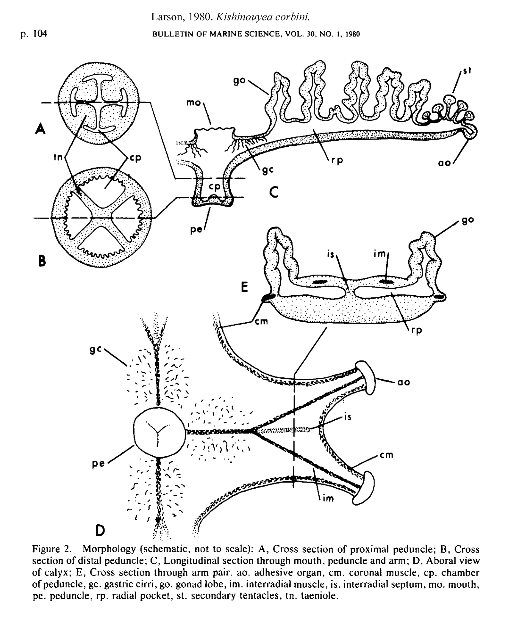
Calyx shallow and well expanded when relaxed, about 15 mm maximum diameter, divided into 4 interradial pairs of arms, resembling a cross which is broadest near its center. Four broadly curved U-shaped perradial notches about twice as deep as the U-or V-shaped interradial notches (Larson, 1980; Grohmann et al., 1999).
Arms each with 5-25 short hollow capitate secondary tentacles on the oral side near the arm tips. Number and size of tentacles increase with increasing calyx size. Tentacles are morphologically alike, although variable in size. The capitate ends of the tentacles are composed of adhesive cells. Primary tentacles lacking in large specimens; only a single 3 mm diameter specimen with primary tentacles was seen. These were similar to the secondary tentacles and were located on the calyx margin between two interradial notches. At the tip of each arm and lying across it, facing aborally, is a broad adhesive pad-like organ which is the result of fusion of several secondary tentacles (Larson, 1980).
Aboral surface is smooth, without ridges or grooves, but is covered with numerous evenly scattered nematocyst warts. The oral surface is smooth but easily folded. Numerous disc-shaped white spots occur on the oral surface near the gonads, mouth and calyx margin. These are vesicles within the mesoglea which are filled with numerous eurytele nematocysts (Larson, 1980).
The manubrium is short and cruciform, the lips highly pleated. The pylorus of the coelenteron contains many (about 200) short gastric cirri. The interradial septa extend from the pylorus almost to the margin, allowing the 4 radial pockets to communicate (Larson, 1980).
Gonads are present in all specimens and are arranged in 8 sinuous adradial bands of closely packed follicles extending from the pyloric region to the tentacles. Along these genital bands, the oral surface is irregularly folded into a number of asymmetrical, highly convoluted, erect lobes which look like small raisins. The number, size, shape and arrangement of these lobes vary from arm to arm and increase with size of the specimen, e.g. 3-mm specimen, 2-3 lobes/arm; 12-mm specimen, 4-8 lobes/arm (Larson, 1980).
The well-developed coronal muscle is separated into 8 U-shaped segments forming a shelf-like extension along the calyx margin. The 4 Y-shaped interradial muscles, also well developed, extend along the interradial septa, bifurcating in the pyloric region and extending to the arm tips (Larson, 1980).
The non-muscular peduncle is short (about 3 mm high in 12-mm specimens), of small diameter (about 1-2 mm), and terminates in a small swollen adhesive pedal disc. It contains a single cruciform chamber, the shape of which is due to the large triangular cross section of each of the four taenioles: only basally within the pedal disc does the stalk become 4-chambered, due to the sudden narrowing of the taenioles into thin radii which unite centrally with a small elevation of the internal surface of the disc. A minute central pit is sometimes present exteriorly on the disc, but no canal arises from it (Larson, 1980).
In life, the coloration is highly variable, but green and reddish brown predominate, which makes them accurate mimics of the Sargassum to which they were attached. The gonads have white with brown, dark green or reddish brown pigmentation along the ridges of the nodular lobes. The gastric cirri were translucent reddish-brown tending to pink .There are white spots of nematocyst in the oral surface (calyx margin and gonads) (Larson, 1980; Grohmann et al., 1999).
Specimens are deposited at the National Museum of Natural History, Washington, D.C., USA (Larson, 1980); and at Cnidarian Laboratory Collection of the Universidade Federal do Rio de Janeiro, Brazil (Grohmann et al., 1999).